(September 2004)
I would like to discuss some of the less common gastrointestinal (GI) diseases of the ferret in this article. Although some of these GI diseases my be uncommon or rare, knowing some of the classic signs of these diseases may help you identify if your ferret has one of these illnesses. Diseases that will be addressed in this issue include megaesophagus, proliferative bowel disease, bacterial infections, enterotoxemia, and parasites. Liver disease and Aleutian Disease Virus (ADV) are diseases that can be associated with GI illness, but they will be discussed more thoroughly in future articles.
1. Megaesophagus
Megaesophagus is a disease in which the esophagus is enlarged and does not have normal motility. It is an uncommon disease, although I have seen several cases over the last few years. Unfortunately the cause of megaesophagus is unknown, and identifying ferrets with the disease is very important as the prognosis for ferrets with this disease is generally poor. Accurately diagnosing megaesophagus early on is important, as there are medications and therapies which can slow and potentially reverse the progression of the disease.
A common clinical sign of megaesophagus is a ferret that regurgitates food 5-10 minutes after eating. These ferrets are often distressed while eating and sometimes appear frustrated because they are very hungry but are physically unable to push the food out of their esophagus into their stomach. Ferrets with this disease are often heard making a gurgling sound during or after eating, and some ferrets choke, cough, or stretch their neck out after eating. Ferrets with megaesophagus generally lose weight quickly as they are effectively starving to death.
A diagnosis of megaesophagus is made based on both clinical signs and x-rays. When a ferret is suspected of having the disease, your veterinarian should give your ferret some soft food with a contrast dye, such as barium, and take x-rays. A positive diagnosis is made if one visualizes retained dye in the esophagus and often a dilated esophagus.
Treatment for this disease is generally supportive, especially since the cause is unknown. The management of megaesophagus in ferrets is similar to that in a dog but is usually less successful. Ferrets are fed thinned, high caloric soft food in a vertical position and then held in a vertical position for a period of time. Hills a/d is commonly used, and supplements such as Nutrical, Dyne, or Deliver 2.0 may be added to increase calorie content. An antacid medication, such as Zantac® can result in dramatic improvement, indicating that gastric reflux and secondary inflammation of the esophagus may be a factor in this disease. Sucralfate (Carafate ®) can be used to coat and therefore promote healing of gastric and esophageal ulcers. Drugs that increase GI motility, such as metoclopramide (Reglan) and cisapride (Propulsid®) may be used to help food empty from the stomach better and reduce gastric reflux. Fluid therapy may also be necessary if the ferret is unable to get adequate fluid intake and has become dehydrated.
Although prognosis is often poor in ferrets with megaesophagus, there have been cases that have done quite well long term with proper medical and dietary management. One of the key factors is identifying the disease as early as possible, before it has progressed very far and before the ferret has become too wasted and weak.
2. Proliferative Bowel Disease
Proliferative Bowel Disease (PBD) is a disease that is caused by a bacterium known as Lawsonia intracellularis. This disease causes inflammation and thickening of the small intestine and/or colon. Ferrets suffering from PBD often have severe wasting which is similar to that seen with ECE (discussed previously). However, in contrast to ECE, this disease most frequently affects rapidly growing juvenile ferrets around 10-16 weeks of age. Environmental and nutritional stress factors appear to be important in this disease, as the incidence of this disease appears to have decreased as the quality of care and nutrition of pet ferrets has improved. Although this disease has become less common and is rare in the Pacific Northwest region of the U.S., identifying ferrets with this disease and beginning treatment early on is crucial, as this disease can be potentially fatal.
Ferrets with PBD have chronic diarrhea that can vary in color and consistency. Feces can be dark and liquidy, have streaks of bright red blood, and/or be mucoid with bright green mucus. The fur around the anus and on the tail may be wet and stained with fecal material. Straining and moaning or crying is often observed, as is protrusion of the rectal tissue. Ferrets suffering from this disease are usually very dehydrated and have low blood protein levels. Since ferrets with PBD are often severely debilitated, they are more susceptible to other infectious diseases such as upper respiratory infections and frequently develop gastritis (inflammation of the stomach) and ulcers. Without appropriate treatment, ferrets with PBD can lose half their weight in less than two weeks. As the disease progresses, ferrets with PBD become very weak and sleep most of the time.
Diagnosis of PBD is usually made based on clinical signs and a ferret being the appropriate age. Your veterinarian may be able to feel the thickened bowel tissue, especially if the colon is involved. Intestinal biopsies and histopathologic examination of the tissue can confirm the diagnosis, however surgery is not recommended in very debilitated animals.
Treatment of this disease includes an antibiotic (chloramphenicol), fluid therapy, nutritional support, and other necessary supportive care. One should make sure that the ferret is housed in a clean, sanitary environment and that the ferret is on an appropriate, high quality diet. Severe damage to the intestine from a disease such as PBD could potentially initiate or result in secondary problems such as inflammatory bowel disease. Therefore, a ferret that has suffered from any severe GI disease such as PBD or ECE should be monitored carefully, both clinically and with appropriate diagnostics such as blood work, throughout its life.
3. Bacterial Infections
Except for Helicobacter which is somewhat commonly observed in pet ferrets, ferrets are relatively resistant to other bacterial diseases of the GI tract. In addition to the bacterium that causes Proliferative Bowel Disease (Lawsonia Intracellularis), there are some bacterial disease that are rarely seen in ferrets such as Salmonellosis and Mycobacteriosis.
Salmonellosis is a contagious disease seen in many species. Ferrets may be exposed to Salmonella organisms by feeding uncooked meat, poultry, and meat by-products. It is characterized by bloody diarrhea, fever, and lethargy. Diagnosis is accomplished by collecting multiple fecal samples and isolating the organism. Treatment consists of antibiotics, aggressive supportive care, and shock therapy if necessary.
Mycobacterium bovis and M. avian infections are rare but have been recognized in research and farm ferrets in England, Europe, and New Zealand. Infection may be associated with feeding raw meat and poultry and unpasteurized dairy products. Diagnosis is done by tissue biopsy. Because this disease has the potential to cause severe illness in people (M. bovis and M. tuberculosis cause tuberculosis in humans), treatment is not recommended.
4. Enterotoxemia
Bacterial enterotoxemia is an uncommon but potentially fatal consequence of severe GI disease and disruption of the normal intestinal bacterial flora. This can occur with diseases such as IBD, bacterial overgrowth, ECE, and other GI diseases. A Clostridium species is the most likely bacterial organism responsible for enterotoxemia.
Treatment of enterotoxemia must be aggressive and rapid. A combination of broad spectrum antibiotics such as metronidazole (Flagyl®) and amoxicillin or enrofloxacin (Baytril®) and amoxicillin, IV fluid therapy, a toxin binding agent such as activated charcoal, nutritional supplementation, and supportive care for symptomatic treatment of diarrhea, shock, hypothermia, etc. are necessary. Enterotoxemia is more easily prevented than treated. Therefore appropriate antibiotics and supportive care for any ferret demonstrating significant GI upset can help prevent or minimize the effects of enterotoxemia. If a ferret dies from suspected enterotoxemia, a diagnosis can be confirmed by performing a necropsy and observing large areas of the small intestine which are dark red to black in color.
5. Parasites
GI parasites are generally rare in ferrets in the United States. The two most common parasites observed in pet ferrets include coccidia and Giardia which are both one celled protozoal organisms. Signs of disease can include diarrhea, straining, and sometimes dehydration or weight loss. Coccidiosis is usually observed in stressed, juvenile ferrets. Diagnosis is made from examination of very fresh feces. Treatment involves the use of anti-parasitic drugs and cleaning up the environment to remove feces and prevent re-infection. Ferrets can pick up other parasites such as tapeworms, roundworms, and hookworms. If other pets in the household have been diagnosed with one of these parasites, then the ferret’s feces should also be checked.
Ferrets
(Before December 23, 2018)
11876 SW Beaverton-Hillsdale Highway
Beaverton, OR
97005
Tel: (503) 646-6101
Email Dr. Ramsell
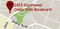
(Starting January 5th, 2019, our new location)
2425 SW Cedar Hills Blvd
Beaverton, OR 97005
Tel: (503) 643-2137
Email Dr. Ramsell

Copyright © 2019 - All Rights Reserved - Northwest Exotic Pet Vet, LLC
Template by Susan's Web Services andOS Templates